Duchenne: electricity and facial expressions
W–064
Duchenne placed electrodes against a face and watched an eyebrow rise or the line of a mouth change. He wanted to separate an expression into the movements of individual muscles. Photography could hold a brief contraction still and make it available for comparison, without relying solely on a drawing or a physician’s memory.
In the mid-1850s, the French physician Guillaume-Benjamin-Amand Duchenne de Boulogne experimented with localized electrical stimulation. Adrien Tournachon, Nadar’s brother, helped with the photographs. They recorded faces from the front and in profile, moved the electrodes and compared the two sides of a face.
One of his principal subjects was an elderly man whom Duchenne described as a former Parisian shoemaker with greatly reduced facial sensitivity. His name has not been established. Duchenne explained that this condition allowed him to stimulate muscles without the usual painful reaction. This was Duchenne’s explanation for choosing the man; it does not tell us what his other subjects experienced.
Online retellings describe these experiments as the torture of restrained patients. Museum catalogues and photographic historian Beatriz Pichel’s study do not give that account of this series. Duchenne stimulated muscular contractions and labelled the resulting expressions terror, pain or joy. A label does not establish what the person felt: an open mouth or a contracted brow could result from the stimulation.
In 1862 he published Mécanisme de la physionomie humaine with a photographic atlas. The work also addressed artists: Duchenne believed that understanding the muscles would help them depict human passions accurately. Its aesthetic section used theatrical subjects, including Lady Macbeth. These were arranged experiments and poses rather than emotions caught by chance.
The photographic plates were products of selection too. Images were cropped, surrounding details masked and photographs combined for comparison. Different prints from the same investigation can therefore retain different boundaries.
In 1868, Charles Darwin showed Duchenne’s photographs to his visitors and recorded the expressions they recognized. Their answers did not always agree. Darwin used this material in The Expression of the Emotions in Man and Animals, published in 1872. The muscular experiments led to another question: how consistently people read faces.
Photographs and documents
1. Electrodes against the face
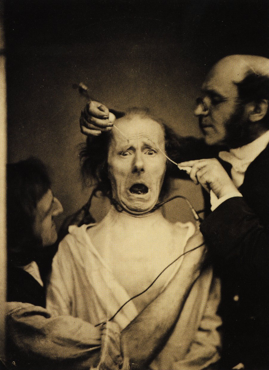
Sources and provenance for frame 1
Wikimedia Commons attributes this photograph to Adrien Tournachon. Its exact day and this copy’s individual museum catalogue number are unestablished. This is the exact submitted screenshot frame. Its source has already removed a narrow edge; WITNESS adds no crop.
The source identifies the historical image as public domain.
File: 902 × 1239 px. Downloaded bytes preserved unchanged; separate uncropped WebP preview. Source file.
Adrien Tournachon (attribution) / Wikimedia CommonsFile on WITNESS · 902 × 12392. Another experiment
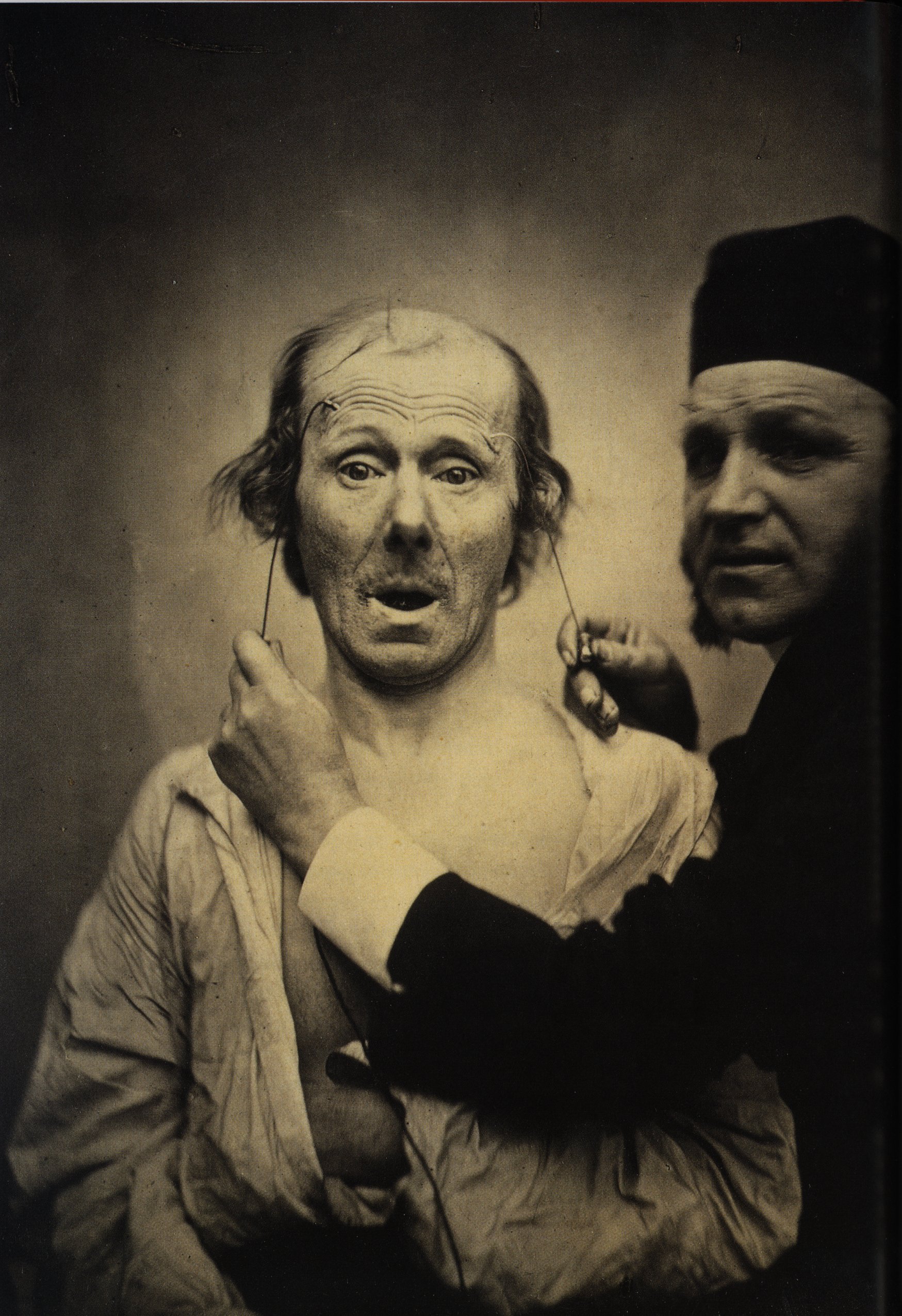
Sources and provenance for frame 2
Wikimedia Commons attributes this photograph to Adrien Tournachon. Its exact day and this copy’s individual museum catalogue number are unestablished.
The source identifies the historical image as public domain.
File: 1749 × 2550 px. Downloaded bytes preserved unchanged; separate uncropped WebP preview. Source file.
Adrien Tournachon (attribution) / Wikimedia CommonsFile on WITNESS · 1749 × 25503. In profile
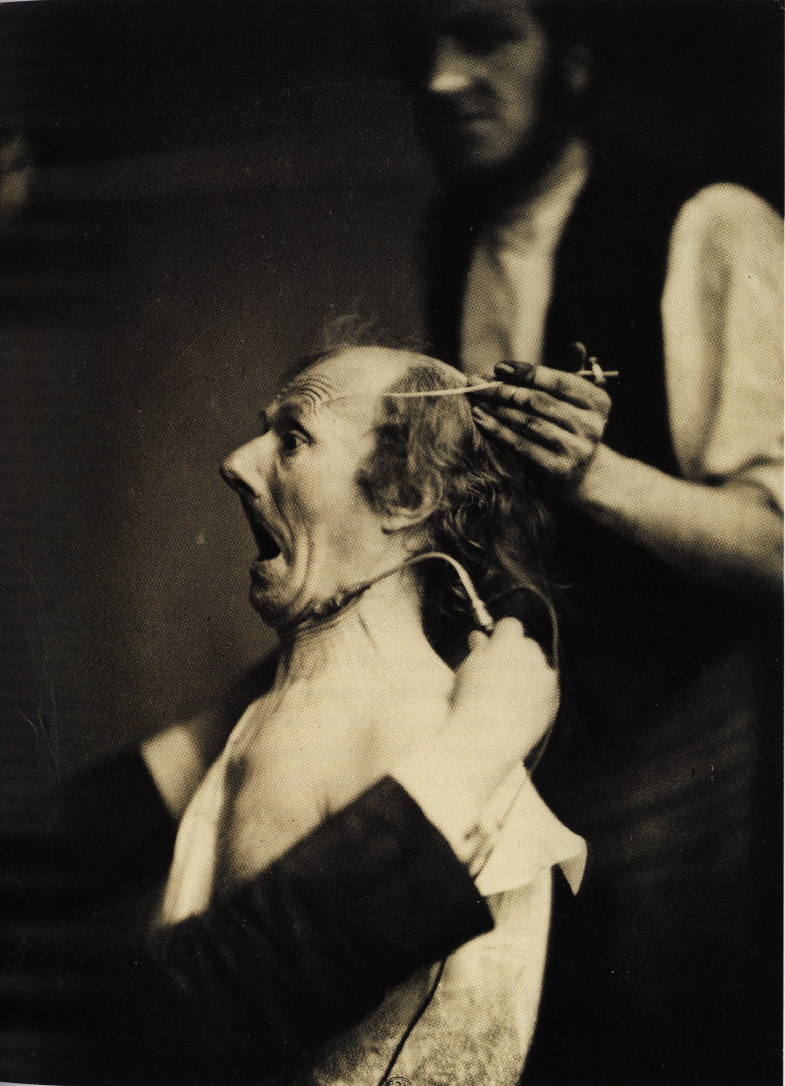
Sources and provenance for frame 3
Wikimedia Commons attributes this photograph to Adrien Tournachon. Its exact day and this copy’s individual museum catalogue number are unestablished.
The source identifies the historical image as public domain.
File: 1551 × 2145 px. Downloaded bytes preserved unchanged; separate uncropped WebP preview. Source file.
Adrien Tournachon (attribution) / Wikimedia CommonsFile on WITNESS · 1551 × 21454. The frontalis muscle
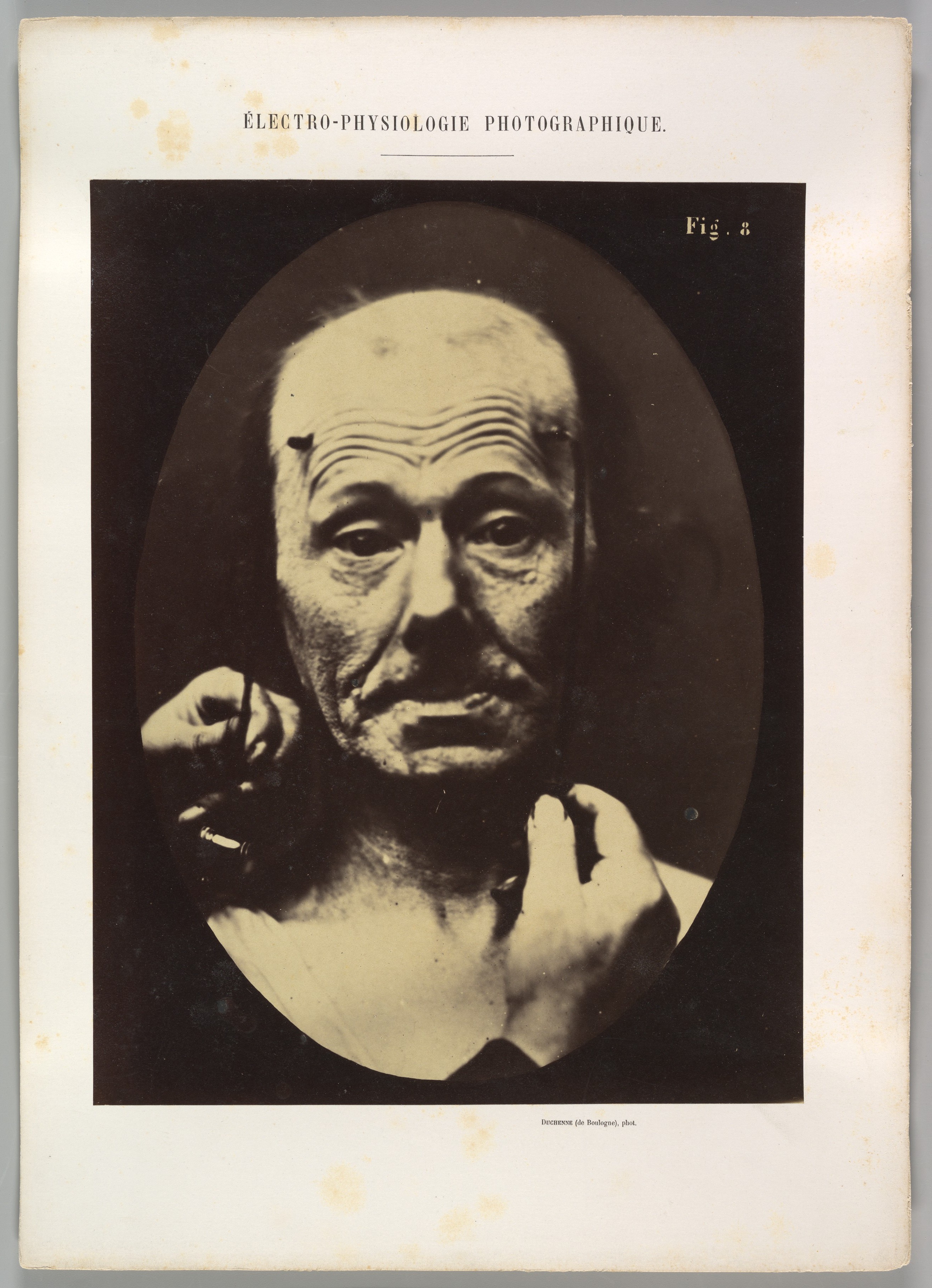
Sources and provenance for frame 4
Accession 2013.173.4. Albumen print from a glass negative; margins and mount retained.
The Metropolitan Museum of Art: Public Domain / Open Access.
File: 2631 × 3634 px. Downloaded bytes preserved unchanged; separate uncropped WebP preview. Source file.
G.-B.-A. Duchenne de Boulogne / Adrien Tournachon / The MetFile on WITNESS · 2631 × 36345. Stimulating the forehead
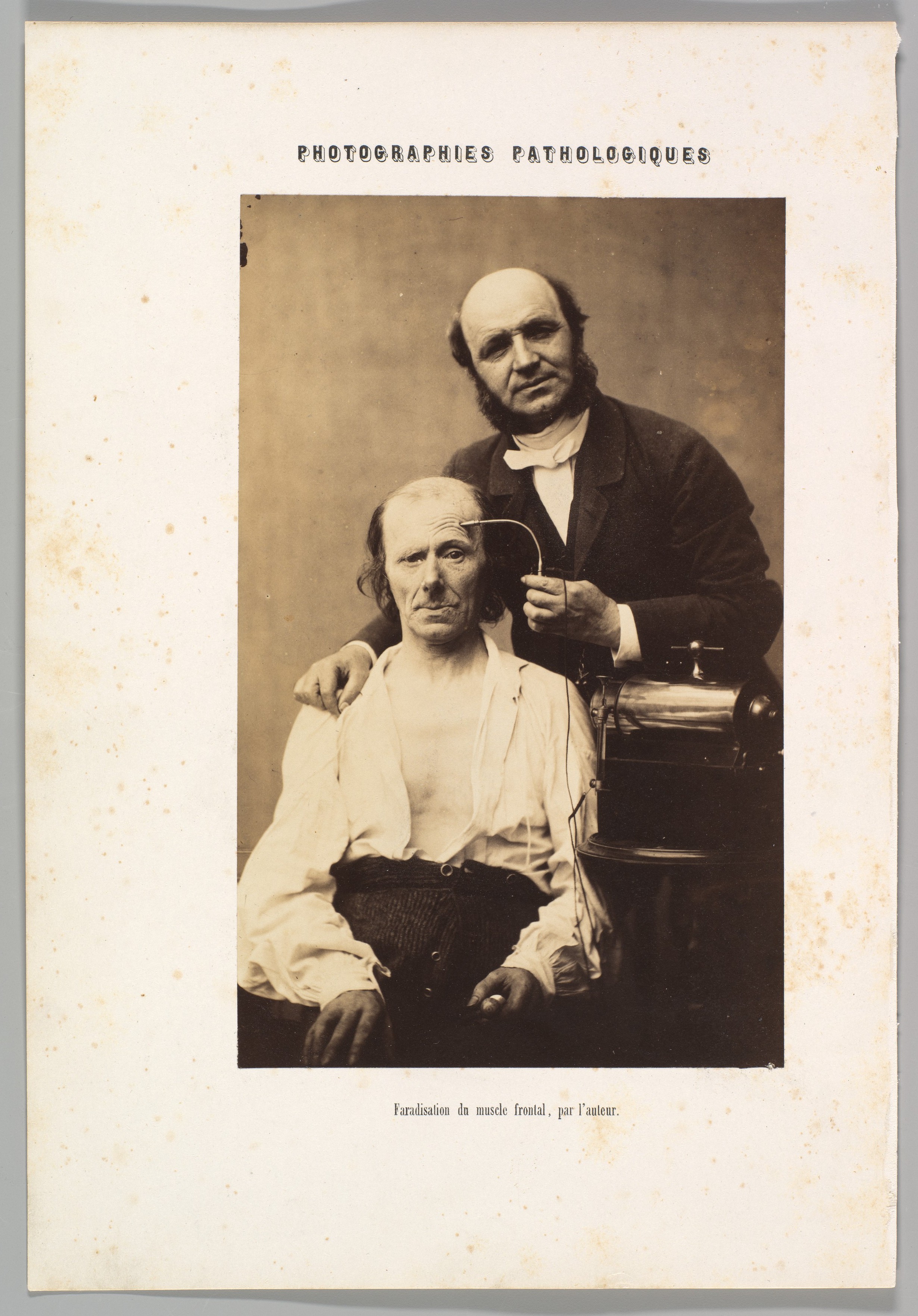
Sources and provenance for frame 5
Accession 2013.229. Albumen print from a glass negative; margins and mount retained.
The Metropolitan Museum of Art: Public Domain / Open Access.
File: 2460 × 3524 px. Downloaded bytes preserved unchanged; separate uncropped WebP preview. Source file.
G.-B.-A. Duchenne de Boulogne / possibly Adrien Tournachon / The MetFile on WITNESS · 2460 × 35246. “Terror”: semiprofile
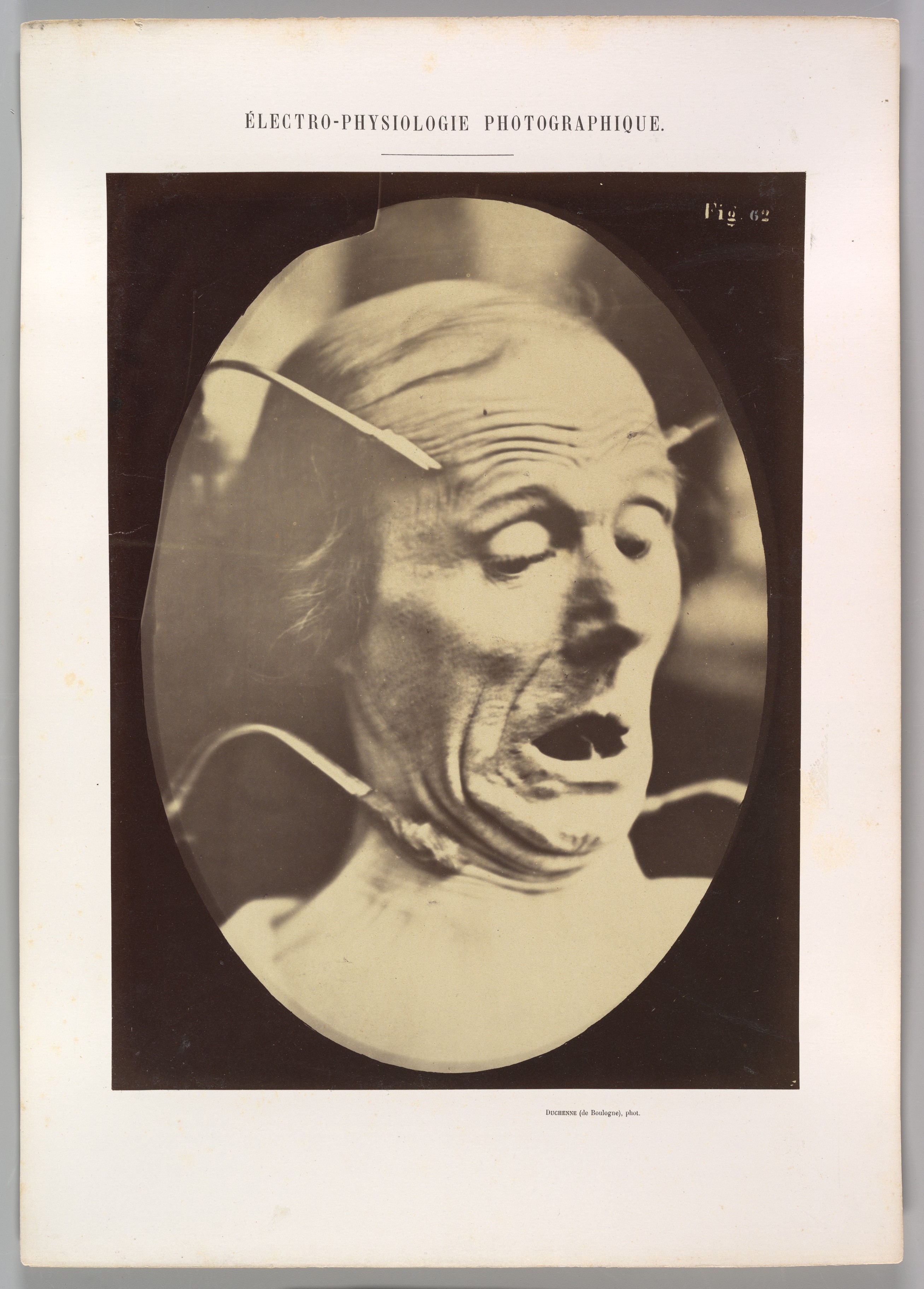
Sources and provenance for frame 6
Accession 2013.173.36. Albumen print from a glass negative; margins and mount retained.
The Metropolitan Museum of Art: Public Domain / Open Access.
File: 2614 × 3645 px. Downloaded bytes preserved unchanged; separate uncropped WebP preview. Source file.
G.-B.-A. Duchenne de Boulogne / Adrien Tournachon / The MetFile on WITNESS · 2614 × 36457. Figure 64
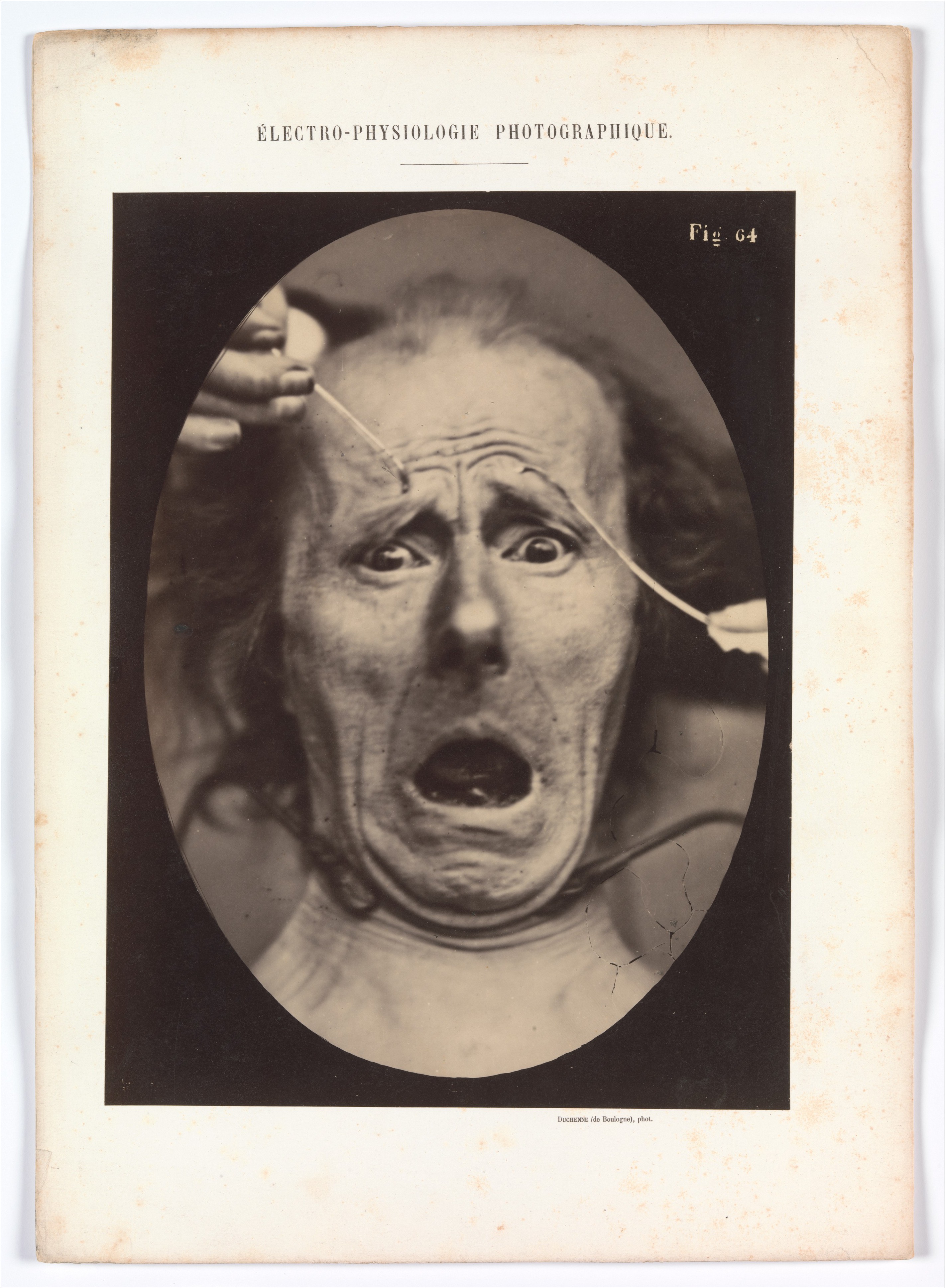
Sources and provenance for frame 7
Accession 2012.140. Albumen print from a glass negative; margins and mount retained.
The Metropolitan Museum of Art: Public Domain / Open Access.
File: 2843 × 3874 px. Downloaded bytes preserved unchanged; separate uncropped WebP preview. Source file.
G.-B.-A. Duchenne de Boulogne / Adrien Tournachon / The MetFile on WITNESS · 2843 × 38748. Figure 65
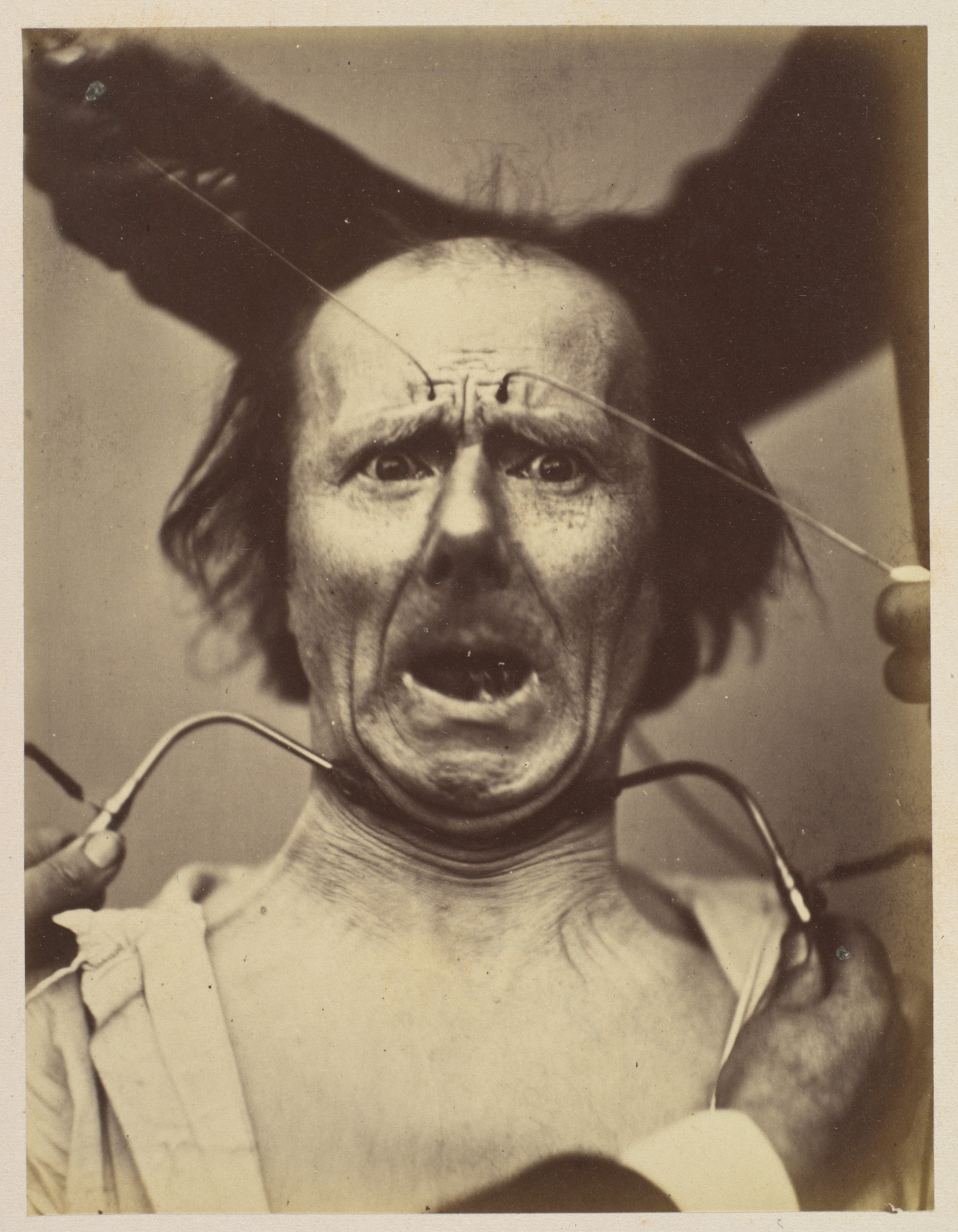
Sources and provenance for frame 8
Accession 1993.248. Albumen print from a glass negative; margins and mount retained.
The Metropolitan Museum of Art: Public Domain / Open Access.
File: 2408 × 3098 px. Downloaded bytes preserved unchanged; separate uncropped WebP preview. Source file.
G.-B.-A. Duchenne de Boulogne / Adrien Tournachon / The MetFile on WITNESS · 2408 × 30989. Lady Macbeth
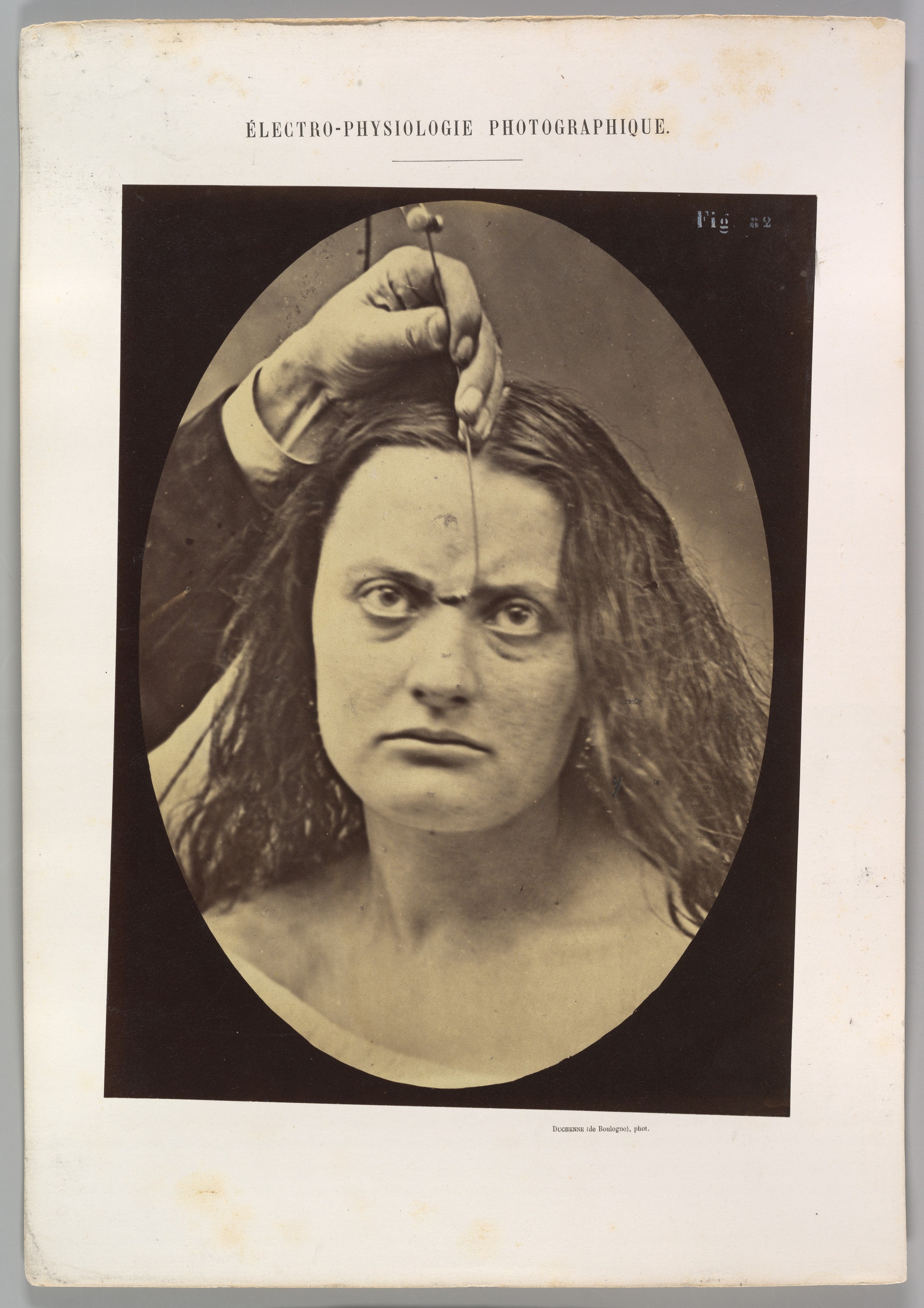
Sources and provenance for frame 9
Accession 2013.173.45. Albumen print from a glass negative; margins and mount retained.
The Metropolitan Museum of Art: Public Domain / Open Access.
File: 2569 × 3636 px. Downloaded bytes preserved unchanged; separate uncropped WebP preview. Source file.
G.-B.-A. Duchenne de Boulogne / Adrien Tournachon / The MetFile on WITNESS · 2569 × 363610. Atlas frontispiece
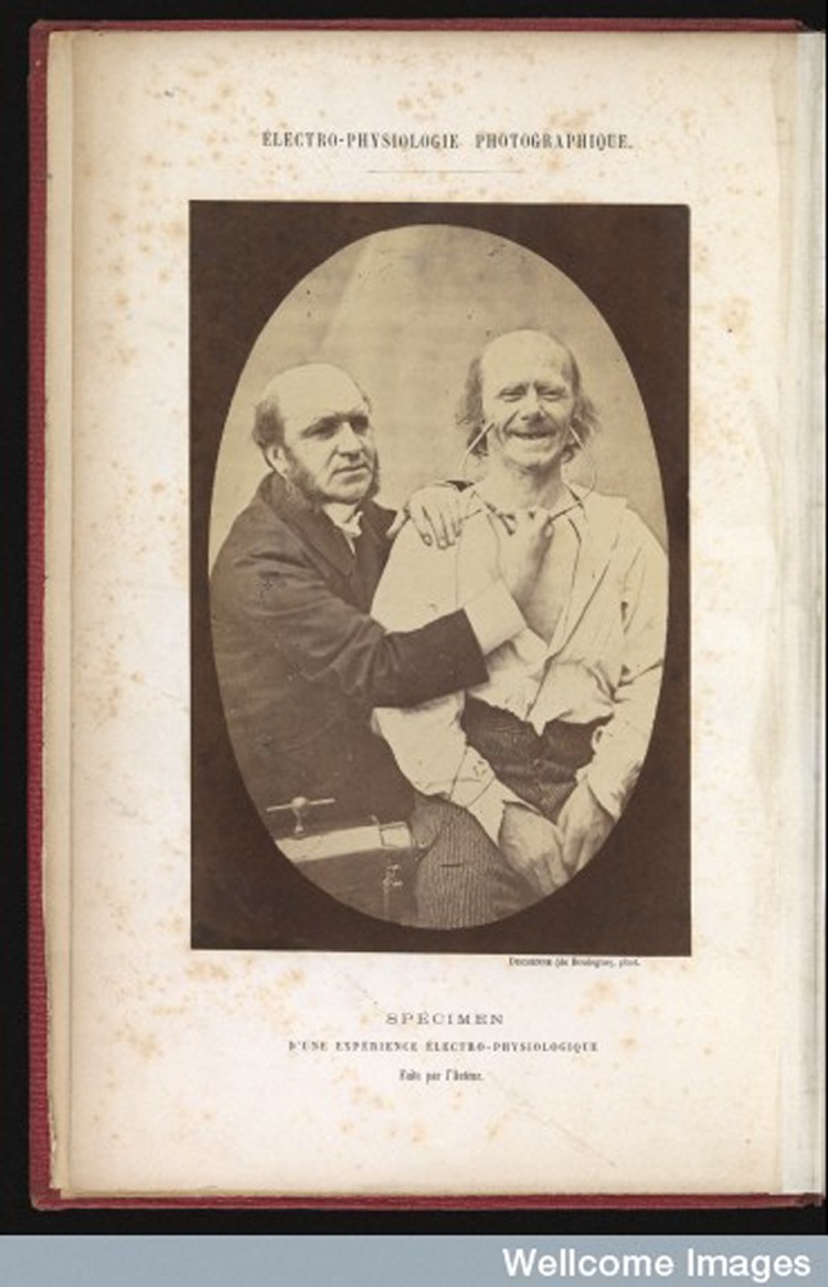
Sources and provenance for frame 10
Available article illustration, not a full museum file. Watermark retained. The larger Wellcome IIIF image could not be obtained because requests timed out.
Reproduction in Beatriz Pichel’s open article (CC BY 3.0); Wellcome Images credit retained. Wellcome’s own digital image record L0077200 specifies CC BY 4.0.
File: 750 × 1165 px. Downloaded bytes preserved unchanged; separate uncropped WebP preview. Source file.
Duchenne de Boulogne / Wellcome ImagesFile on WITNESS · 750 × 1165